
Can Hong Kong PGT Screen for Chromosomal Abnormalities? PGT-A and PGT-SR Technology Explained

Hong Kong PGT can screen for chromosomal abnormalities. PGT-A screens for numerical abnormalities, and PGT-SR screens for structural abnormalities. Hong Kong fertility centers use NGS technology with a detection accuracy exceeding 98%. This article provides a detailed explanation of the principles, procedures, suitable candidates, and precautions for PGT screening of chromosomal abnormalities.
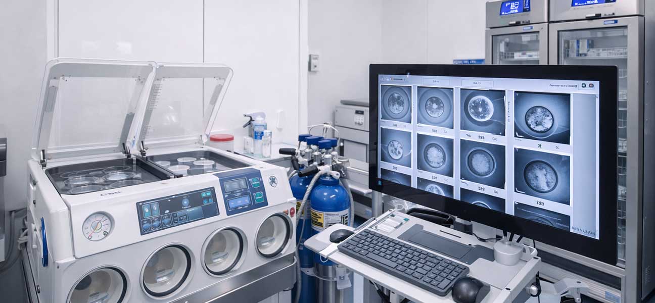
Opening: Direct Answer
Hong Kong PGT can screen for chromosomal abnormalities. PGT-A screens for numerical chromosomal abnormalities (aneuploidy), and PGT-SR screens for structural chromosomal abnormalities (balanced translocations, Robertsonian translocations, inversions, etc.). Hong Kong fertility centers commonly use NGS technology for whole-genome analysis of all 23 pairs of chromosomes, achieving a detection accuracy of over 98%.
Why Can PGT Screen for Chromosomal Abnormalities?
Chromosomal abnormalities are a major cause of embryo implantation failure, early miscarriage, and birth defects. Chromosomal abnormalities are divided into two main categories: numerical abnormalities (e.g., trisomy 21, trisomy 18, trisomy 13, monosomy X, etc.) and structural abnormalities (e.g., balanced translocations, Robertsonian translocations, inversions, deletions, duplications, etc.).
The core principle of PGT technology is: when the embryo develops to the blastocyst stage on day 5-6, 5-10 cells are biopsied from the trophectoderm. Sufficient DNA is obtained through whole genome amplification, and then NGS (Next-Generation Sequencing) or aCGH technology is used to sequence and analyze the DNA, thereby determining the chromosomal status of the embryo.
Technical features of Hong Kong PGT include:
- Uniform use of blastocyst biopsy for more accurate assessment of embryo developmental potential
- NGS technology covering all 23 pairs of chromosomes with a resolution of 5-10 Mb
- Combined with vitrified frozen embryo transfer strategies to avoid the impact of ovarian stimulation cycles on the endometrium
- Laboratories generally hold international certifications such as CAP/CLIA, with mature quality control systems
Actual Process of Hong Kong PGT for Screening Chromosomal Abnormalities
From the start of ovarian stimulation to obtaining PGT results, the complete cycle typically takes 4-6 weeks. The specific steps are shown in the table below:
| Stage | Content | Time |
|---|---|---|
| Pre-assessment | Karyotype analysis for both partners, genetic counseling, fertility assessment | 1-2 weeks |
| Ovarian Stimulation | Medication for stimulation, follicle monitoring, egg retrieval surgery | 10-14 days |
| Fertilization & Culture | ICSI fertilization, embryo culture to blastocyst | 5-6 days |
| Blastocyst Biopsy | Biopsy of 5-10 trophectoderm cells | 1 day |
| Whole Genome Amplification | DNA amplification of biopsied cells | 1-2 days |
| NGS Sequencing Analysis | Sequencing and data analysis of amplified products | 7-14 days |
| Embryo Freezing | Immediate vitrification of embryos after biopsy | 1 day |
| Result Interpretation | Genetic counselor interprets report, selects transferable embryos | 1-2 days |
| Transfer | Select euploid or balanced embryos for transfer | 1 day |
Most Hong Kong fertility centers use "spent embryo DNA analysis" combined with NGS technology, which can provide PGT-A reports within 2-3 weeks. PGT-SR may take longer, 3-4 weeks, due to the need for breakpoint analysis or family linkage analysis.
Clinical Perspectives of Reproductive Doctors on PGT Screening for Chromosomal Abnormalities
In clinical decision-making, PGT-A is primarily used for screening chromosomal abnormalities in the following groups:
- Female age ≥ 35 years
- History of recurrent miscarriage (≥ 2 times)
- History of recurrent implantation failure (≥ 3 times)
- Severe male factor infertility
- History of pregnancy with chromosomal abnormalities
The indications for PGT-SR are more specific:
- One partner is a carrier of a balanced translocation
- One partner is a carrier of a Robertsonian translocation
- One partner is a carrier of an inversion
- One partner has other confirmed structural chromosomal abnormalities
From clinical data, the aneuploidy rate in embryos from women under 35 is about 20%-30%, and it can exceed 60%-70% in women over 40. PGT-A can effectively screen for euploid embryos, increasing the clinical pregnancy rate per single transfer to 50%-65% while reducing the miscarriage rate to below 10%. However, PGT is not suitable for everyone—in cases of diminished ovarian reserve, low oocyte yield, or limited embryo numbers, PGT carries the risk of having no embryos available for transfer, which requires thorough counseling.
Differences in PGT Strategies by Age Group
Age is the most critical factor affecting the rate of embryonic chromosomal abnormalities, and PGT strategies vary significantly across different age groups:
| Age Group | Embryo Aneuploidy Rate (Reference) | Clinical PGT Recommendation |
|---|---|---|
| < 35 years | Approx. 20%-30% | PGT may not be necessary without clear genetic indications |
| 35-37 years | Approx. 30%-40% | PGT-A is recommended |
| 38-40 years | Approx. 40%-55% | PGT-A is strongly recommended |
| 41-42 years | Approx. 55%-70% | PGT-A is strongly recommended; risk of no embryos must be discussed |
| > 42 years | > 70% | PGT-A is recommended, but expectations for oocyte yield should be clearly communicated |
Older women benefit more significantly from PGT-A—with a marked reduction in miscarriage rates and more efficient transfer cycles. However, the absolute number of euploid embryos obtained decreases with age, and some cycles may be canceled due to the absence of normal euploid embryos. Clinicians need to make individualized judgments based on AMH, antral follicle count (AFC), and previous stimulation history.
Differences Between Hong Kong and Mainland China PGT
There are several differences between Hong Kong and Mainland China regarding PGT technology for chromosomal abnormality screening:
| Dimension | Hong Kong | Mainland China |
|---|---|---|
| History of Technology Application | PGT technology applied for over 15 years | PGT technology applied for about 10 years |
| Legal Regulation | Clear legal framework, well-defined indications | Strict medical indication approval required; PGT-SR/M needs reporting |
| Testing Scope | Comprehensive coverage of PGT-A/SR/M, can detect > 300 monogenic diseases | PGT-A relatively lenient; PGT-SR/M requires approval with limited disease panel |
| Laboratory Certification | Commonly hold international certifications like CAP/CLIA | Primarily domestic certifications; some centers have international accreditation |
| Embryo Biopsy Method | All use blastocyst biopsy | Some centers still perform cleavage-stage biopsy (day 3) |
| Reporting Time | 2-3 weeks (PGT-A), 3-4 weeks (PGT-SR) | 2-4 weeks (PGT-A), 4-6 weeks (PGT-SR) |
| Ability to Detect Complex Abnormalities | Extensive experience with complex translocations, microdeletions, mitochondrial diseases, etc. | Complex abnormalities may require referral or external testing |
Hong Kong has accumulated richer clinical data and laboratory experience in the PGT field, with stronger capabilities for detecting complex chromosomal abnormalities (e.g., triple translocations, cryptic translocations, uniparental disomy, etc.). Additionally, Hong Kong's genetic counseling system is more comprehensive, allowing patients to receive more informed decision-making support.
Easily Overlooked Details
In the clinical practice of PGT screening for chromosomal abnormalities, the following details are often overlooked but have significant implications for result interpretation and transfer decisions:
1. Mosaicism Issues
Embryos may contain a mixture of normal and abnormal cells (mosaicism). PGT biopsy only samples 5-10 trophectoderm cells, which may not fully represent the genetic status of the entire embryo. Embryos with low-level mosaicism (< 30%) may still develop into healthy fetuses, but this requires comprehensive assessment based on embryo morphology score and patient condition. High-level mosaicism (> 70%) is generally not recommended for transfer.
2. Biopsy Timing and Cell Source
Blastocyst biopsy is superior to cleavage-stage biopsy because blastocysts have more cells, and embryos tolerate the biopsy better. However, the genetic concordance between the inner cell mass (which develops into the fetus) and the trophectoderm (which develops into the placenta) is about 95%-97%, meaning there is a 3%-5% discordance rate. In rare cases, PGT results may differ from the actual fetal karyotype.
3. Detection Resolution Limitations
NGS technology has high accuracy for detecting chromosomal abnormalities larger than 5 Mb, but may miss microdeletions/microduplications smaller than 5 Mb. If a specific microdeletion syndrome is clinically suspected (e.g., 22q11.2 deletion, 15q11.2 deletion, etc.), targeted probes or dedicated SNP arrays should be used for supplementary testing.
4. De Novo Mutations and Unclear Breakpoints
PGT-SR is accurate for detecting structural abnormalities with known breakpoints. However, if the abnormality is a de novo mutation or has unclear breakpoints, the standard PGT-SR protocol may not be sufficient. In such cases, whole-genome sequencing or customized probes for family analysis may be required.
5. Embryo Freeze-Thaw Risks
After biopsy, embryos need to be vitrified and stored while waiting for PGT results. The survival rate after freeze-thaw in Hong Kong centers is about 95%-98%, but there is still a small risk of embryo damage during the thawing process. For patients with a limited number of embryos, this risk should be considered in advance.
Management of Special Situations
Balanced Translocation Carriers
Among embryos produced by balanced translocation carriers, approximately 50%-70% have unbalanced chromosomes. PGT-SR can screen for balanced or completely normal embryos for transfer. Hong Kong's PGT-SR typically uses breakpoint detection combined with SNP linkage analysis, which can distinguish between balanced translocation carriers and completely normal embryos—this is particularly important for families who wish for the next generation not to carry the translocation.
Robertsonian Translocation Carriers
Robertsonian translocations mainly involve chromosomes 13, 14, 15, 21, and 22. Among embryos produced by carriers, about 30%-50% are normal or balanced. PGT-SR combined with NGS can effectively screen for these, but dedicated analysis strategies need to be designed based on the specific translocation type.
Clinical Decision-Making for Mosaic Embryos
For embryos with PGT results indicating mosaicism, clinical management should consider the following factors:
- Mosaic ratio: Low-level mosaicism (< 30%) may be considered for transfer; high-level (> 70%) is generally not recommended.
- Specific chromosome involved: Mosaicism involving certain chromosomes (e.g., 1, 3, 4, etc.) has greater clinical significance.
- Embryo morphology score: Mosaic embryos of good quality are more likely to be considered for transfer.
- Patient age and number of embryos: For older patients with few embryos, a low-level mosaic embryo may be the only option, requiring thorough informed consent.
Repeated PGT Cycles with No Euploid Embryos
If, after multiple PGT cycles, there are still no normal euploid embryos, a reassessment is needed:
- Analysis of the source of chromosomal abnormalities in sperm or eggs (e.g., sperm FISH, polar body analysis)
- Consideration of egg or sperm donation
- Whether to attempt transfer of embryos without PGT (requires assessment of age and miscarriage risk)
- Possibility of occult germline mosaicism or de novo mutations
Fertility centers in Hong Kong typically organize a multidisciplinary consultation (involving reproductive doctors, genetic counselors, and embryologists) after 2-3 PGT cycles to formulate a subsequent plan for the patient.
0 comments